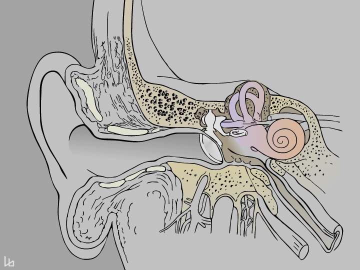
The ear is divided in three major parts, outer, middle and inner ear. The snail-like structure called „cochlea“ harbours all cells necessary to perceive sounds. In deafness, these cells are severely damaged and the auditory nerve receives no information.
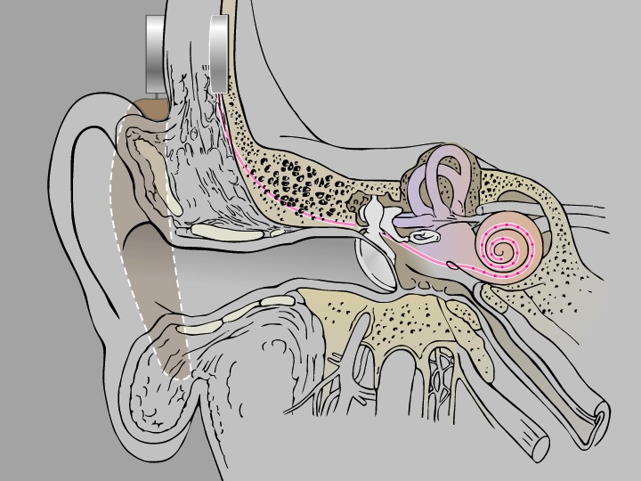
A cochlear implant restores hearing in deaf patients. It consists of an externally worn speech processor and an implanted stimulator receiver equipped with an electrode array (pink). The intracochlear electrodes directly stimulate auditory nerve fibres situated at a distance of 200 micrometers.
The cochlea contains fluid-filled spaces, which do not allow auditory nerve fibres to pass. To enable outgrowth of nerve fibres towards the electrode array, a matrix has to be injected, which will guide and support the neurons towards the target.
Cochlea with implanted electrode array.
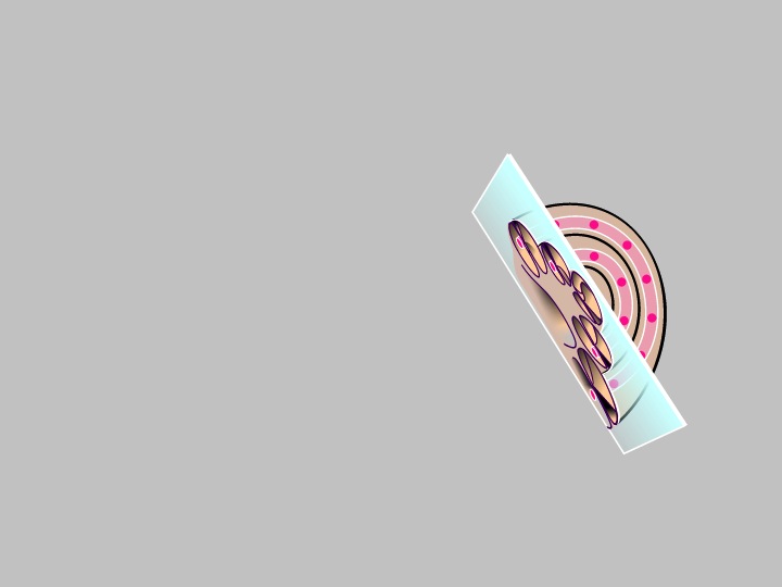
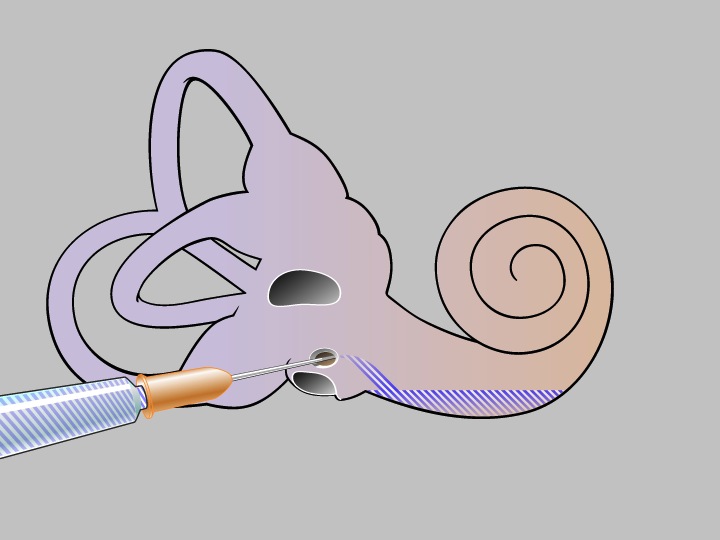
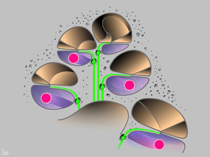
The following images show a cross-section of the cochlea (mid-modiolar section).
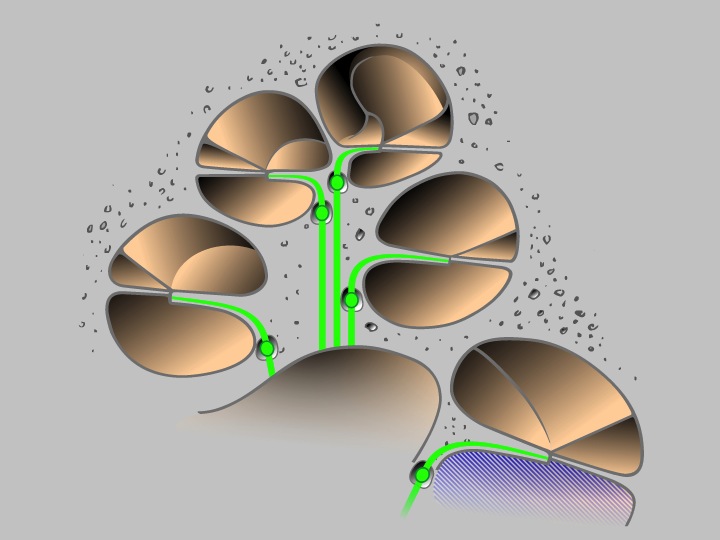
The cochlea in a mid-modiolar section with the fluid-filled cochlear turns and the auditory neurons (green). The nanomatrix is inserted into the scala tympani of the basal turn (blue).
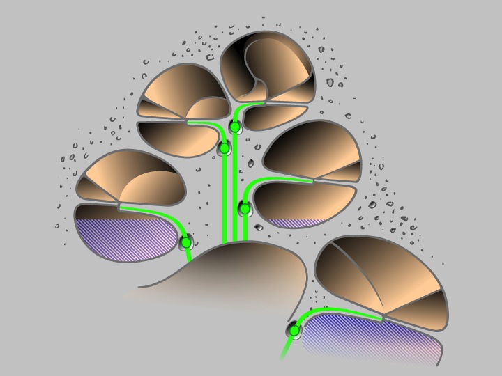
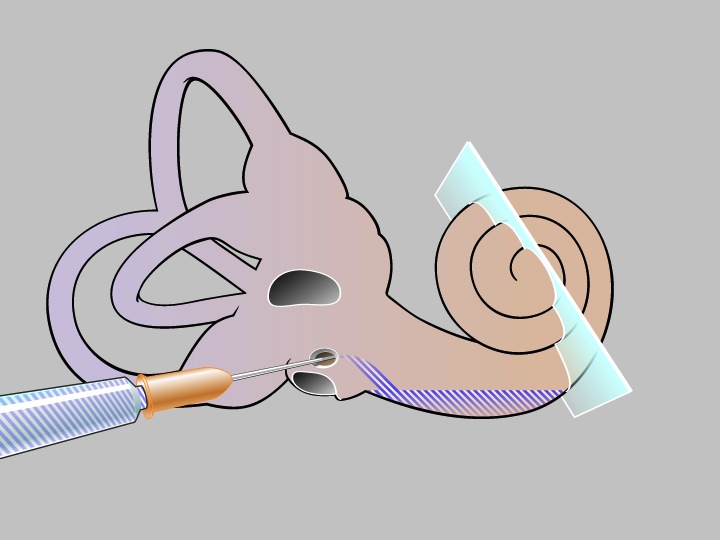
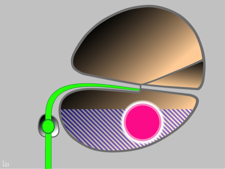
The scala tympani is gradually filled from the base towards the apex with the nanomatrix (blue).
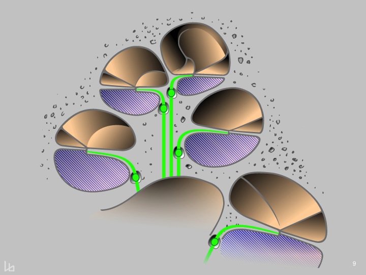
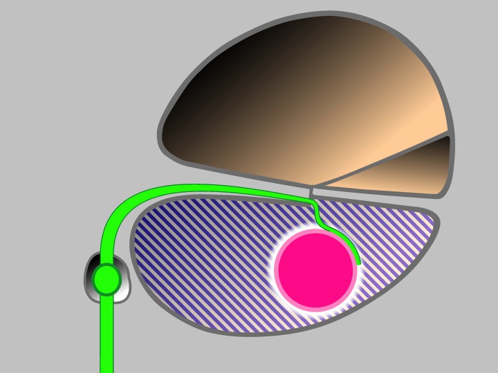
The entire scala tympani is now filled with the nanomatrix (blue), which should enable outgrowth of auditory nerve fibres (green).
The matrix-filled scala tympani with the nano-CI electrode array in situ (pink). The distance from the auditory nerve fibres to the electrode array surface ranges from 50 – 200 micrometers.
This is a zoom into a cross-section of one turn of the spiral. The gap between the electrode array (pink) and the symbolized auditory nerve fibre (green) leads to overlappling electrical fields, because several hundreds of neurons are stimulated simultaneously from one electrode contact. Therefore the resolution of sounds of the cochlear implant is lower compared to the natural situation.
The main goal of the NANOCI-project is to create a gapless interface between the auditory nerve fibres (green) and the electrode array (pink). To overcome the fluid-filled scala-tympani, a nano-matrix loaded with growth- and guidance factors (blue-white) is injected prior to cochlear implantation. The gapless interface would have several theoretical advantages, such as higher resolution of neural stimulation and lower energy consumption.
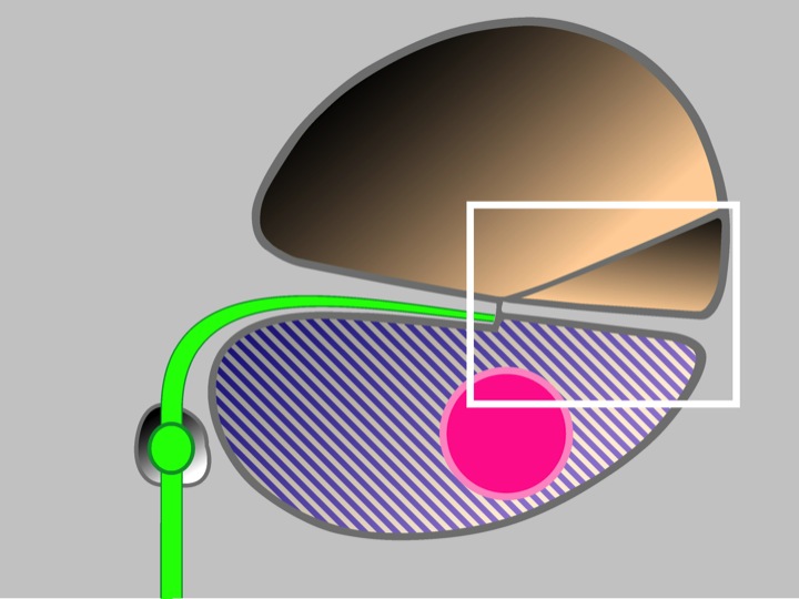
The following images show a zoom into the region where the auditory nerve fibre establishes contact.
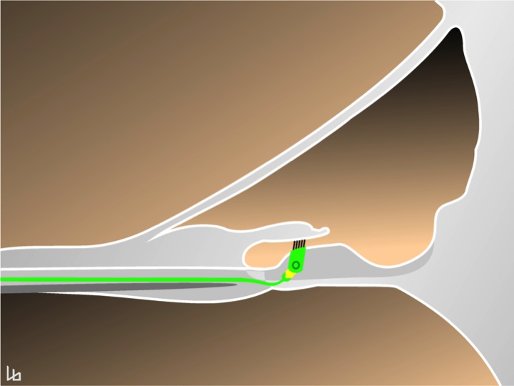
In the normal hearing ear, the auditory nerve fibre (green) gaplessly contacts the inner hair cell via a synapse on the cell body (yellow).
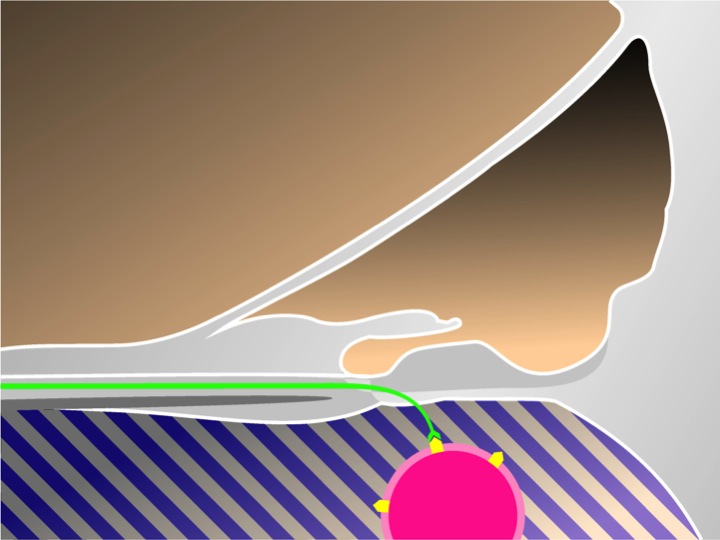
The dendrite of the auditory neuron in the deaf ear (without hair cell) is guided through the neurotrophic nano-matrix (blue-white) and stably locked on the nanostructured electrode array surface (pink with yellow symbols) via a gapless interface, mimicking the natural situation.